

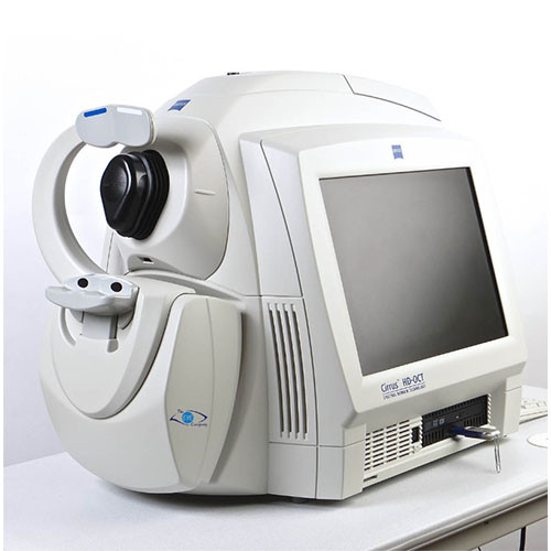
Zeiss Cirrus 4000 OCT (Pre-Owned)
Brand: Zeiss
SKU: OTUZE4000
Price:
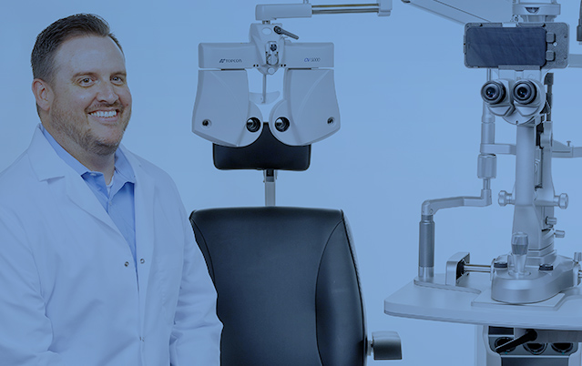
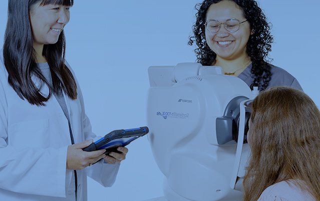
The pre-owned Zeiss Cirrus HD-OCT 4000 enables examination of the posterior and anterior of the eye at an extremely fine spatial scale, without surgical biopsy or even any contact with the eye.
Free
MSRP:
Employing the advanced imaging technology of spectral domain optical coherence tomography, the Cirrus 4000 acquires OCT data about 70 times faster (27,000 vs. 400 A-scans per second) and with better resolution (5 μm vs. ~10 μm axial resolution in tissue), compared to first-generation OCT technology. Cirrus acquires whole cubes of OCT image data, composed of hundreds of line scans, in about the same time as Stratus acquires a six-line scan. You can view these data cubes in three planes, or through three dimensions, giving you access to an extensive amount of retinal image data in one scan.
Includes warranty covering parts, labor, and shipping. All pre-owned (used) inventory from Lombart is completely refurbished, tested, and re-calibrated by trained certified technicians. Please allow up to 4 weeks for delivery.
Product may differ from image based on availability. Please contact us for details. All pre-owned and demo products are subject to availability.
FEATURES
- Unprecedented Visualization of Anatomical Details
- HD Layer Maps
- HD Enhanced Raster Scan
- Cirrus HD-OCT Scan Engine
- Correlation Between OCT Scan and Fundus Image
- Superior Image Data
- Designed for efficiency
- Small footprint and integrated design are ideal for crowded or busy practice
- 90 degree orientation facilitates observation of patient throughout exam
- Advanced optics aid in the examination of patients with cataracts. Dilation is not required even for pupils as small as 2.5 mm
- Mouse Driven Alignment™ delivers superior image capture and analysis in just a few clicks, resulting in reduced chair time for the patient
- Auto Patient Recall™ assures patient position and instrument setting are repeated from previous visit
SPECIFICATIONS
Technical data
- Axial resolution: 5 μm (in tissue)
- Transverse resolution: 15 μm (in tissue)
- Scan speed: 27,000 A-scans per second
- A-scan depth: 2.0 mm (in tissue), 1024 points
- Optical source: superluminescent diode (SLD), 840 nm
Fundus Imaging
- Line scanning ophthalmoscope (LSO)
- Live during scanning
- Transverse resolution: 25 μm (in tissue)
- Optical source: superluminescent diode (SLD), 750 nm
- Field of view: 36° x 30°
Scan Patterns
- Macular Cube 200 x 200 Combo: 200 horizontal scan lines comprised of 200 A-scans
- Macular Cube 512 x 128 Combo: 128 horizontal scan lines comprised of 512 A-scans
- 5 Line Raster: 4096 A-scans per B-Scan (adjustable length, spacing and orientation)
Focus Adjustment Range
- –20D to +20D (diopters)
Fixation
- Internal and external
Pupil Size Requirement
- ≤ 2.0 mm (≥ 3.0 mm optimal for LSO)
Computer
- Windows® X 7 Ultimate
- High-performance multi-core processor
- Internal storage: > 80,000 scans
- CD-RW, DVD-ROM drive
- Integrated 15” color flat panel display
Electrical
- 100–120V~, 50/60Hz, 5A 220–240V~, 50/60Hz, 2.5A
Related Products
A Division of Advancing Eyecare™
© 2024 Lombart Healthcare All Rights Reserved.
Account Information
Get to Know Lombart
Lombart Healthcare is committed to keeping our site accessible to everyone. We welcome feedback on ways to improve the site’s accessibility so it is easy for everyone to navigate.
Free shipping is only available for online orders in the continental United States.
Certain exclusions apply.
Exclusions include, but are not limited, to the following products:
Acuity Systems & Projectors, Chair & Stand Accessories, Autorefractors, Lensmeters, Keratometers, Portable Slit Lamps, Stools, Tables, Tonometers, Trial Lens Sets.
- Exam Lane
- Pre-Test
- Diagnostic/Imaging
- Treatment & Surgical
- Lab & Dispensing
- Lens Edgers
- Supplies & Accessories
- Pre-Owned
- All Pre-Owned Equipment
- Pre-Owned Autorefractor/Keratometers
- Pre-Owned Biometers
- Pre-Owned BIOs
- Pre-Owned Chairs & Stands
- Pre-Owned Edgers
- Pre-Owned Lasers
- Pre-Owned Lensmeters
- Pre-Owned Manual Keratometers
- Pre-Owned OCTs
- Pre-Owned Projectors
- Pre-Owned Refractors & Phoroptors
- Pre-Owned Retinal Cameras
- Pre-Owned Slit Lamps
- Pre-Owned Tonometers
- Pre-Owned Ultrasounds
- Pre-Owned Visual Field Perimeters